Venous Ulcer vs. Arterial Ulcer: How to Tell Which Type of Leg Wound You Have
Published 2026-09-11

There is an open sore on your lower leg, and two people have already given you two different answers. The pharmacist said "probably circulation." Your neighbor said her husband had "the same thing" and needed a stent.
Both could be right, because two very different diseases open wounds on the lower leg. One comes from veins that cannot push blood up. The other comes from arteries that cannot bring blood down.
Vein specialists at Vein Center Doctor sort out that question every week for patients across New York and New Jersey. The answer changes everything about treatment.
Venous Ulcer vs. Arterial Ulcer: What Is the Difference?
A venous ulcer is a shallow, irregular wound near the inner ankle caused by chronic venous insufficiency, where failed valves let blood pool and damage the skin. An arterial ulcer is a deep, punched-out foot or shin wound caused by narrowed arteries starving the tissue.
The distinction is not academic. Venous ulcers heal with compression and treatment of the refluxing vein. Arterial ulcers need the blocked artery reopened, and compression can make them worse.
Roughly seven of every ten leg ulcers are venous, so the odds favor a vein problem. The remaining cases, arterial and diabetic wounds, are the ones a specialist must rule out before any bandage goes on.
How to Tell If a Leg Ulcer Is Venous or Arterial
Each type of ulcer leaves a signature in where it sits, how it looks, and how it hurts. The table below compares venous, arterial, and diabetic (neuropathic) ulcers across the features a specialist checks first:
| Feature | Venous Ulcer | Arterial Ulcer | Diabetic (Neuropathic) Ulcer |
|---|---|---|---|
| Location | Inner ankle and lower calf (gaiter zone) | Toes, heel, outer ankle, shin, bony points | Sole of the foot, ball of the foot, under toes |
| Wound bed | Shallow, red, moist, often weeping | Deep, pale or gray, dry, sometimes black tissue | Pink or red, often deep, surrounded by callus |
| Edges | Irregular, sloping, ill-defined | Sharp, "punched-out," well-defined | Round, rimmed with thick callus |
| Pain pattern | Aching and heaviness, worse standing, better elevated | Severe, worse when the leg is raised or in bed at night, eased by dangling the foot | Often painless because nerves are damaged |
| Surrounding skin | Brown staining, swelling, eczema, hardened skin | Shiny, thin, hairless, pale or bluish, cool | Dry, cracked, thick callus, foot deformity |
| Pulses and temperature | Pulses present, leg warm | Pulses weak or absent, foot cold | Pulses usually present, foot warm but numb |
Two of those rows do most of the work. Location tells you which vessel system is failing, because venous pressure lands at the inner ankle while arterial starvation hits the toes and heel first.
Pain finishes the job. A wound that feels better when you put your feet up is almost always venous. A wound that wakes you at night until you hang your leg off the bed is arterial until proven otherwise.
The arterial ulcer vs venous ulcer location rule has exceptions, and mixed disease is common in patients over 65. That is why the table is a starting point, not a diagnosis.
Why Veins Cause One Ulcer and Arteries Cause the Other
Leg veins carry blood back to the heart against gravity. One-way valves stop it from falling back down between heartbeats.
When those valves fail, blood refluxes and pools in the lower leg. That pooling drives sustained pressure, called venous hypertension, into the tiny vessels at the ankle.
The pressure forces fluid, iron, and inflammatory cells into the skin. Skin under that kind of stress stiffens, stains brown, and eventually breaks open at the slightest knock.
This is the pathway behind chronic venous insufficiency, and untreated varicose veins are its most visible warning sign. The ulcer is the end stage of a vein problem that has usually been building for years.
Arterial ulcers start from the opposite direction. Peripheral arterial disease narrows the arteries with plaque, so less oxygen-rich blood reaches the foot.
Tissue that cannot get enough oxygen cannot repair itself. A small pressure point or a tight shoe rub becomes a wound that will not close.
Smoking, diabetes, high blood pressure, and high cholesterol drive that plaque. The same risk factors cause heart attacks and strokes, which is why an arterial ulcer is a whole-body warning, not just a foot problem.
Diabetic foot ulcers sit in a third category. Nerve damage removes the pain signal, so a patient walks on a blister until it becomes a crater.
Diabetic foot ulcer vs venous ulcer is often the easier call. The diabetic wound is on the sole, painless, and ringed with callus. The venous wound is at the ankle, achy, and surrounded by stained skin.
Duplex Ultrasound and the Ankle-Brachial Index
No specialist should diagnose a leg ulcer by eye alone. Two painless tests settle the venous-versus-arterial question in a single visit.
Duplex ultrasound maps the veins. The scan shows which valves have failed, how far blood is refluxing, and whether a clot is hiding in a deep vein.
Vein Center Doctor uses this scan as the first step of every ulcer workup. It converts a vague "circulation problem" into a specific vein that can be treated.
The ankle-brachial index (ABI) checks the arteries. A technician measures blood pressure at the ankle and at the arm, then divides one by the other.
The number reads like this:
- 1.0 to 1.4 is normal arterial inflow.
- 0.9 or below indicates peripheral arterial disease, and the lower the value, the worse the blockage.
- Below 0.5 signals severe arterial disease, and wounds at this level rarely heal without the artery being reopened.
- Above 1.4 usually means calcified, stiff arteries, common in diabetes, and the reading cannot be trusted without further testing.
Here is why the ABI matters so much for compression. Compression bandages squeeze the leg to push pooled venous blood upward, which is exactly what a venous ulcer needs.
Put that same squeeze on a leg whose arteries are already struggling to deliver blood, and the bandage can shut off the little inflow that remains. The result can be tissue death, worsening ulcers, and in the worst cases amputation.
That is why a vein specialist confirms an ABI of 0.8 or higher before ordering full-strength compression. An ulcer that hurts more inside a bandage needs the bandage off and the arteries checked.
How Vein Center Doctor Treats a Venous Ulcer
Once ultrasound confirms venous reflux and the ABI clears the arteries, the healing plan attacks the pressure from two sides.
Graduated compression therapy comes first. Multilayer bandages or prescribed stockings lower venous pressure, control swelling, and give the wound bed the conditions it needs to fill in. Worn consistently, compression alone closes many venous ulcers within a few months.
Closing the refluxing vein stops the ulcer from coming back. Radiofrequency ablation seals the failing saphenous vein with targeted heat through a thin catheter.
VenaSeal closes the same vein with medical adhesive and no heat. It suits patients who want to skip compression stockings after the procedure.
Smaller feeder veins around the ulcer get ultrasound-guided sclerotherapy, an injection that collapses the vein so blood reroutes through healthy channels. Every procedure is outpatient, performed by the team under Medical Director Dr. Rahul Sood, and most patients walk out the same day.
Arterial ulcers are a different specialty. If the ABI shows significant arterial disease, Vein Center Doctor refers the patient to a vascular surgeon or interventional specialist. That team handles arterial imaging and, where needed, angioplasty, stenting, or bypass.
Treating an arterial ulcer with vein procedures does not work, and honest vein specialists say so plainly.
Mixed ulcers, where both systems are failing, get the arteries addressed first and modified compression only once inflow is safe. For a deeper look at what stalls healing once the diagnosis is right, see why a leg ulcer won't heal.
When a Leg Ulcer Needs Urgent Care
Most leg ulcers can wait for a scheduled specialist visit. Some cannot.
Go to an emergency department or call your doctor the same day for:
- A foot or toe that has turned black, blue, or white and cold.
- Severe rest pain in the foot that started or worsened in the past few days.
- Fever, chills, or a red border spreading rapidly around the wound.
- Foul-smelling drainage, exposed bone or tendon, or a sudden jump in pain.
- A wound that appeared after a fall or injury and is bleeding heavily.
The first two signs point to critical limb ischemia, an arterial emergency where hours matter. The next two point to deep infection, which can spread into bone.
For everything else, book a vein screening within the next week or two. Brown staining, itching, and hardening skin above the ankle are the early warning signs of a venous leg ulcer.
Catching them before the skin opens is the easiest win in vein care.
Find Out Which Ulcer You Have With a Free Vein Screening
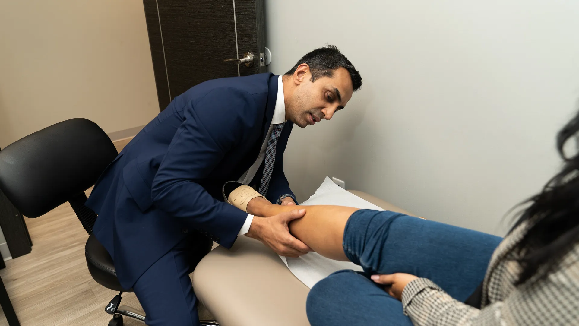
Vein Center Doctor offers free vein screenings at 12 locations across New York and New Jersey. A board-certified specialist examines the wound, maps your veins with duplex ultrasound, and checks arterial flow.
You leave knowing whether the problem is venous, arterial, or both, and which specialist should treat it.
Schedule your free leg ulcer screening today or call (914) 274-4412. Same-week appointments are available at offices from Ardsley to Edison.