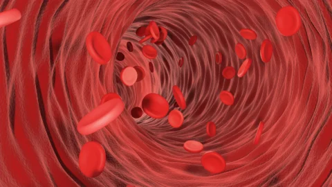
Research about a disease is always geared towards getting to know more about the disease, even to its molecular level. This is because knowing the intricacies of a certain disease is tantamount to determining ways to address and manage it.
So can chronic venous insufficiency affect deep veins? Yes, because chronic venous insufficiency occurs as a consequence of defects in valves, a structure that most veins possess. Further proving this is the fact that a common complication of the CVI is deep vein thrombosis.
The Difference Between Superficial and Deep Veins
Superficial and deep veins are 2 of the different types of veins. They are so named because of their location. As the name suggests, superficial veins are near the body’s surface while deep veins are located far from the surface. Superficial veins also don’t have a corresponding artery in contrast to deep veins which have a corresponding one.
Other types of veins include the pulmonary veins which transport oxygen-rich blood from the lungs and to the heart and the systemic veins which transport oxygen-poor blood from all the other body parts back to the heart.
How Chronic Venous Insufficiency Occurs
Chronic venous insufficiency is a vascular disease characterized by the failure of blood to return to the heart from the legs, causing the blood to pool in the veins. This is a consequence of reflux, obstruction, or a combination of the two.
Venous reflux occurs in an interplay of different factors, but the main causes include the following:
- the incompetence of the venous valve;
- inflammatory reactions in the vessel wall;
- changes in the dynamics of blood flow; and
- venous hypertension.
On the other hand, venous obstruction which causes a limitation in blood flow that leads to impairments in calf muscle pump function may be caused by the following:
- deep vein thrombosis;
- venous stenosis;
- extrinsic compression.
Clinical Manifestations of Chronic Venous Insufficiency
The presentation of chronic venous insufficiency may well be seen through inspection of its most prominent signs and symptoms. These signs and symptoms may further be understood better through a standardized scheme called the CEAP Classification Score complemented by the Venous Clinical Severity Score.
1) Signs and Symptoms of Chronic Venous Insufficiency
Chronic venous insufficiency may present with a wide spectrum of conditions, including dilated veins, subjective symptoms, inflamed skin changes and skin irritation, and venous ulcers. Specifically, CVI may present with the following signs and symptoms:
- Dilated leg veins – Dilated leg veins may be called differently based on their sizes. They may be called telangiectasias or spider veins when they’re < 3 mm in size, reticular veins when they’re 1 to 3 mm in size, and varicose veins when their sizes are > 3 mm.
- Lower extremity edema – Edema is the scientific term for swelling due to fluid build-up. They start from the perimalleolar region and then travel up to the legs when they occur in the lower extremities.
- Subjective symptoms – Subjective symptoms that include aching, heaviness, and restless leg syndrome are suggestive of chronic venous insufficiency especially when they’re aggravated when exposed to heat, with a preponderance to time, and when they can be improved when you rest your legs or elevate them.
- Skin changes – Skin changes include hyperpigmentation or the darkening of the skin due to blood leakage in the ankle region, eczema or stasis dermatitis characterized by blistering and scaling, lipodermatosclerosis, and atrophie blanche (literally white atrophy).
- Venous stasis ulcer – Venous stasis ulcers are chronic, non-healing wounds associated with chronic venous insufficiency that occurs due to venous hypertension.
2) The CEAP Classification Score
The CEAP classification score is the widely used classification in describing and standardizing chronic venous insufficiency outcomes. It’s a classification scheme with an acronym that stands for clinical signs for C, etiology for E, anatomical distribution for A, and pathophysiology for P.
They can further be classified into clinical classes, with increasing severity of disease and clinical manifestations as features for each class, in that there are no inspectable signs of venous disease present for Class 0 while skin changes with active ulceration are characteristic for Class 6.
3) Venous Clinical Severity Score
The venous clinical severity score is a system used to further quantify and standardize the reporting of the severity of chronic venous insufficiency. It serves to complement the results of the CEAP classification score for a better description of CVI for clinical use.
This scoring system is particularly helpful for the determination of the patient’s response to treatment and is recommendable as a scoring system for use in clinical practice.
Complications of Chronic Venous Insufficiency

Complications of chronic venous insufficiency are blood clots that occur in the superficial veins and the deep veins. These are as follows:
- Superficial thrombophlebitis – Superficial thrombophlebitis is the inflammation and clotting that occurs in the vein immediately found under the skin. It’s characterized by severe leg pain but usually isn’t serious. It goes away on its own usually within 2 to 6 weeks and is responsive to treatment.
- Venous thromboembolism – Venous thromboembolism is an encompassing term for deep vein thrombosis (DVT) and pulmonary embolism (PE). It occurs when a blood clot causes obstruction in blood flow (DVT) or travels up to the lungs (PE).
- Post-thrombotic syndrome – PTS is the disease condition that occurs after a patient has contracted DVT, causing non-relieving pain, swelling, and other symptoms in the legs. It happens due to the damage in the venous valves and vein walls caused by DVT.
How Chronic Venous Insufficiency is Diagnosed
Chronic venous insufficiency may be diagnosed with careful history taking and physical examination, and imaging techniques that may be non-invasive testing procedures or invasive.
1) History Taking and Physical Examination
History taking is done by interviewing the patient to assess the severity of the disease in accordance with the patient’s perception. It may also be used to determine the risk factors that a patient possesses that have an association with CVI, which include the following:
- older age
- sedentary lifestyle
- obesity
- pregnancy
- phlebitis
- previous leg injury
- occupational or environmental factors like prolonged standing or sitting
- a medical history and family history of cardiovascular risk factors and diseases like deep venous thrombosis and heart failure
Meanwhile, a physical exam entails the inspection of the legs for dilated veins and other irregularities like dilated tortuous veins. Other clinical manifestations like skin changes and edema are observed and assessed for their severity as well.
Superficial and deep venous reflux may also be differentiated during physical examination, in a maneuver called the classic tourniquet test where applying a tourniquet or manual compression is done. In this maneuver, superficial venous reflux is suspected when it takes more than 20 seconds for the varicose veins to dilate, while a rapid dilation is suspected for deep venous reflux.
2) Non-Invasive Testing
Non-invasive testing serves to assist and complement the physical exam findings. Included under this classification are the imaging techniques that don’t require an injection of a contrast medium or any other invasive technique toward the vein that the healthcare provider would want to visualize.
Examples of diagnostic testing procedures under this category include:
- Venous duplex testing
- Air plethysmography
- Computed tomography or magnetic resonance venography
- Photoplethysmography
- Strain gauge plethysmography
- Foot volumetry
3) Invasive Testing
Invasive testing procedures involve direct visualization of the target veins by injection of a contrast medium, catheterization of the target veins, and inserting a needle that’s connected to a transducer in the dorsal foot vein. Examples of invasive testing procedures include:
- Contrast venography
- Intravascular ultrasound
- Ambulatory venous pressure monitoring
Treatment of Chronic Venous Insufficiency
Strategies in treating chronic venous insufficiency include conservative treatments, under which are pharmacologic treatments for symptomatic relief and lifestyle modification including regular exercise, and interventional treatment option that includes surgical repair like vein stripping, and outpatient vein treatments.
Moreover, signs and symptoms that may warrant hospitalization in patients with CVI include non-responding cellulitis and massive weeping edema, hypotension, abnormal laboratory findings like low serum bicarbonate levels, and severe disease indicative of soft tissue infection.
Vein Center Doctor: Offering State-of-the-Art Outpatient Vein Treatments
You don’t have to worry about whether or not chronic venous insufficiency affects deep veins. At Vein Center Doctor, we’re committed to addressing your blood vessel problems with our state-of-the-art outpatient vein treatments that can reach even the deepest veins. They can bring back your legs with damaged veins to a much healthier state.
1) Radiofrequency Ablation
Radiofrequency ablation is a kind of thermal ablation that entails the delivery of radiofrequency energy through a heat-tipped catheter that’s advanced up to the saphenofemoral junction. It’s performed under general or local anesthesia and guided by ultrasound. This procedure works by obliterating the damaged vein by inflicting injury to the target vein that causes the constriction of this vein.
This procedure, however, can’t be done in small veins where catheterization is impossible and in patients with residual thrombosis. Moreover, complications associated with this procedure include burns, paresthesias, bruising, infection, and superficial thrombophlebitis.
2) Endovenous Laser Treatment
Endovenous laser ablation involves also the catheterization of the target vein and advancing it up to the saphenofemoral junction. The laser-tipped catheter is used to deliver laser energy that’s also used to obliterate the vein. This procedure is also done under general or local anesthesia and is also under ultrasound guidance.
Also similar to radiofrequency ablation, endovenous thermal ablation also can’t be done in small veins where catheterization is impossible. It also has its complications, which include bruising, and hyperpigmentation, among others.
3) Sclerotherapy
Sclerotherapy is the procedure of choice when it comes to small veins. It involves injecting a chemical agent called sclerosant into the target vein to cause its closure due to the irritation of the blood vessel wall. Sclerotherapy may be in liquid or foam form, with foam being the more effective but more painful option. Typical sclerosants used include a hypertonic solution of saline and sodium tetradecyl sulfate.
This procedure can’t be done in patients that are pregnant or breastfeeding, with known septal defects or patent pulmonary ovale, and allergy to the sclerosant used. SIde effects associated with this procedure include hyperpigmentation, superficial phlebitis, deep vein thrombosis, and skin necrosis.
4) VenaSeal Closure System
The VenaSeal closure system offers the option to maximize patient comfort by not requiring anesthesia, sclerosants, and the use of compression therapy after the procedure. This treatment entails the delivery of 0.1 cc of the VenaSeal adhesive through a special catheter for 3 seconds then compressing the area for 3 minutes.
This will be repeated until all of the target vein is covered. It requires the washing of the area first with saline solution and the guidance of ultrasound in the determination of the access site and the delivery of the adhesive.
Side effects associated with this procedure include allergic reactions against the VenaSeal adhesive, arteriovenous fistula, and bleeding in the access site.
5) Compression Therapy
The first-line treatment is compression therapy for patients with chronic venous insufficiency and lower extremity venous ulcers. It entails the delivery of an external compression that’s graded through the use of elastic compression stockings and pneumatic compression devices.
This graded compression serves to oppose the hydrostatic pressures brought by CVI that cause edema and other complications like venous leg ulcers.
This technique, however, has associated issues like problems in patient compliance and the lack of standard pressure even when the compression garment is wrapped around by a healthcare professional. For best results, seek the help of a vein care professional.
Get World-Class Outpatient Vein Care at Vein Center Doctor
Chronic venous insufficiency can occur in both superficial and deep veins because most veins have valves and one of the mechanisms by which CVI occurs is having incompetent valves. Similarly, both a complication and a risk factor of CVI is deep vein thrombosis, which is a blood clot that forms in the deep veins.
At Vein Center Doctor, we’re committed to bringing our patients the best outpatient vein care from superficial venous conditions to even the deepest vein issues. Our innovative medical devices combined with the expertise of our team headed by Dr. Rahul Sood ensure procedures are performed with accuracy, precision, and patient-centered care. Contact us today at 1-862-500-4747 for your free consultation.


