Can Pelvic Venous Insufficiency Cause Lower Abdominal Pain?
Published 2022-05-24

Pain in women is one of the most common causes of gynecological referrals, especially when concerning their menstrual period. However, many conditions associated with this clinical presentation are often underdiagnosed, often documented only as chronic pain of unclear etiology. One of these underdiagnosed conditions is pelvic venous insufficiency.
So can pelvic venous insufficiency cause lower abdominal pain? Pelvic venous insufficiency is a disease characterized by both chronic and dull pelvic and abdominal pain. It's common among female patients of child-bearing age (premenopausal women) and those who have given birth to many offspring. It's also associated with coexisting depression and anxiety.
Pelvic Venous Insufficiency As A Cause Of Lower Abdominal Pain
Pelvic venous insufficiency or pelvic congestion syndrome is a vascular disease referring to the pathological process where the retrograde flow of blood in the pelvic and ovarian veins, which results in the pooling of blood into these veins and their consequent dilatation. This then manifests as pelvic varicose veins and pain in the lower abdomen.
The pathophysiology leading to this disease is multifactorial, and factors that may contribute to the exacerbation of this venous disease include the following:
- A family history of varicose veins;
- Hormonal influences and the disturbance in hormonal regulation;
- Previously undergoing pelvic surgery;
- Having a retroverted uterus or a uterus where the tip is directed towards the rectum instead of the belly;
- Multiple pregnancies;
- The nutcracker syndrome or the sequelae of conditions manifesting with left flank pain and microscopic hematuria due to the compression of the left renal vein;
- The renal vein having a high blood flow which is a normal characteristic of its physiology;
- The hemodynamic changes associated with pregnancy such as high blood flow in the arteries and in the uterine veins, leading to venous reflux.
Characteristics of Lower Abdominal Pain Caused By Pelvic Venous Insufficiency
The most pressing sign of pelvic venous insufficiency is chronic or recurrent abdominal pain (that is, the pain has lasted for at least 6 months), although acute and severe pain may also occur. The pain may be exacerbated by standing and the hormonal changes that occur during the menstrual cycle, especially during the premenstrual period. The pain associated with pelvic venous insufficiency includes the following characteristics:
Localization
whole abdomenlower abdomenflanksback
Side
leftright
Character
constantcolickystabbingdull
Other Signs and Symptoms of Pelvic Venous Insufficiency
Some signs of pelvic venous insufficiency may be gynecological, urological, and cardiovascular. The presence of these symptoms along with a physical examination finding of ovarian point tenderness strongly suggests the diagnosis of pelvic venous insufficiency, with 94% sensitivity and 77% specificity.
- dyspareunia (pain during urination)
- hematuria (blood in the urine)
- dysuria (decreased frequency of urination)
- urinary frequency
- urgency
- varicose veins which may be seen in the lower extremities, vulva, and anus
Depression and anxiety are also commonly seen together with the occurrence of pelvic venous insufficiency, which may be due to the length of time it takes before a diagnosis of pelvic venous insufficiency can be made. It may also be due to the secretion of vasodilator substances in response to the stress from the ovarian vein dilation, which can also play a role in the regulation of emotion and stress.
Diagnostic Imaging Findings That Suggest Pelvic Venous Insufficiency
Clinical investigation of pelvic venous insufficiency may be done through diagnostic imaging modalities such as Doppler ultrasound, computed tomography scan, magnetic resonance imaging, transcatheter venography, endocavitary examination, and phlebography.
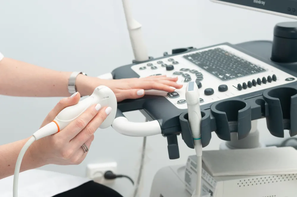
1) Doppler Ultrasound
Doppler ultrasound is considered to be the first-line diagnostic modality that aids in confirming a clinical suspicion of pelvic venous insufficiency. It's used to determine the presence of reflux by looking at sites opposite the anatomic zones.
If a female patient did not undergo pelvic surgery, the origin of the varicosities may be determined by classifying them into saphenous or non-saphenous. Saphenous varicosities are further defined in the involvement of the saphenofemoral junction and posterior communicating vein while non-saphenous varicosities are concluded to be of pelvic origin.
Three sonographic criteria must be met in order to ascertain the diagnosis of pelvic venous insufficiency:
- pelvic vein dilatation, with the diameter being greater than 4 mm
- slow-moving blood flow (less than 3 centimeters per second)
- arcuate vein dilatation (this communicates with pelvic varicosities)
2) Computed Tomography Scan and Magnetic Resonance Imaging
The use of computed tomographic scan or magnetic resonance imaging can help exclude other causes of chronic pelvic pain, which may not be detected using ultrasound. These include endometriosis, fibroids, and adenomyosis, among others.
They can also detect the presence of a varicocele ovarian vein incontinence, which may be asymptomatic and can't also be seen using doppler ultrasound. This is due to their high tissue contrast and spatial resolution.
MRI is also considered more advantageous than CT scan, especially when used with gadolinium contrast medium because of lesser radiation, ability to identify the direction of blood flow, and better sensitivity and specificity.
3) Transcatheter Venography
Transcatheter venography is performed when the diagnosis of pelvic venous insufficiency hasn't been adequately supported by the previously discussed non-invasive diagnostic methods. Typical findings suggesting pelvic venous insufficiency include the following:
- ovarian vein dilation (moderate if at 5-8 mm diameter and severe if with a diameter of greater than 8 mm)
- retrograde ovarian reflux
- uterine venous engorgement
- cross filling of pelvic veins from the filling of vulvovaginal or thigh varicosities
Furthermore, a score of 5 or more in the venographic scoring system strongly suggests pelvic venous insufficiency, with 91% sensitivity and 89% specificity. The components of this scoring system are as follows:
- maximum diameter of the ovarian vein
- time to the disappearance of contrast material
- degree of congestion
4) Endocavitary Examination
Using a 5 to 7.5 MHz endocavitary transducer, visualization of veins may be done, such as the uterine and lateral vaginal venous plexuses, the adnexal veins, and the transuterine venous passages.
Dilation of these veins may be interpreted when a sonographic finding of anechoic cords is observed. Pelvic venous insufficiency may be considered upon doing this procedure when the following observations are seen:
- vein with a diameter of more than 9 mm
- vein with a sinuous appearance
- abundance of veins
5) Phlebography
Phlebography may be considered both diagnostic and therapeutic, done when Doppler ultrasound confirmation of utero-ovarian vein incontinence was done. This procedure is done under local anesthesia where catheters and guidewires are used to assess the diameter of veins, the size of varicoceles, and the length of time the contrast medium disappears. There are 2 approaches that may be done under this procedure: brachial or femoral approaches.
Treatment of Pelvic Venous Insufficiency
Upon confirmation of the diagnosis of pelvic venous insufficiency, its medical management may be done through different procedures available, such as:
- Medical options - The principle behind the medical option of treating pelvic venous insufficiency stems from the idea of treating this venous disease as if it's also an ovarian dysfunction. Hormonal therapies are included under this treatment option, which includes medroxyprogesterone acetate (MPA) and goserelin, and an implant of etonogestrel.
- Surgical options - Surgical options that may be done for pelvic venous insufficiency include the laparoscopic ligation of the ovarian veins and hysterectomy.
- Interventional options - Interventional options for the treatment of pelvic venous insufficiency entail the transcatheter embolization (or blocking) of ovarian varices using coils as an embolic agent and performing a follow-up to check for the improvement of symptoms.
Expert Outpatient Vein Treatments at Vein Center Doctor
Studies have shown that at least 50% of women with pelvic venous insufficiency also suffer from reflux of the deep veins of the lower extremities. A medical history of varicose veins also often warrants looking for the medical history of pelvic venous insufficiency. It's therefore also important to address the signs and symptoms of pelvic venous insufficiency as well as chronic venous insufficiency.
At Vein Center Doctor, we offer expert outpatient vein treatments that we perform with the patient's safety and comfort as the top priority for our clinical practice. Quality medical care in our clinic includes radiofrequency ablation, endovenous laser treatment, sclerotherapy, VenaSeal closure system, and compression therapy, which are all proven to be effective treatment methods for vascular conditions without the need for surgical treatments such as vein stripping.
1) Radiofrequency Ablation
Radiofrequency ablation is a type of interventional treatment for chronic venous insufficiency. It entails the delivery of radiofrequency energy, usually in the greater saphenous vein, through an endovenous electrode to obliterate the blood vessels that are already incompetent. This results in the redistribution of blood flow from the veins with reflux to normal, healthy veins.
This process involves the use of a catheter with a heating tip, which is passed through the vein under general or local anesthesia up to the saphenofemoral junction. However, this can't be done in patients with smaller veins that the cannula can't pass through. Patients with residual venous thrombosis also can't undergo this procedure.
Side effects associated with this procedure include burns, bruising, and infection. Observation of the patient for the occurrence of deep vein thrombosis after the procedure is also an important aspect to be done.
2) Endovenous Laser Treatment
Endovenous laser treatment also employs the use of heat to destroy the endothelium, the inner lining of the blood vessels, to close off the incompetent veins. Under local anesthesia and through the visualization of ultrasound, a laser-tipped catheter is passed through the greater saphenous vein up to the saphenofemoral junction to deliver a laser beam and form a scar tissue that closes off the vein. Through this closing off of the affected vein, the flow of blood is then redirected towards healthier veins.
The effects of endovenous laser treatment are less rapidly seen compared to that of radiofrequency ablation. It's also not applicable to varicose veins with small diameters where cannulation is impossible. It can't also be done on patients with residual thrombosis. Depending on the strength of the laser beam used, the severity of pain and other complications such as hyperpigmentation and bruising may increase.
3) Sclerotherapy
Sclerotherapy entails the administration of chemicals called sclerosing agents or sclerosants via the Tessari method. Typical sclerosants used are sodium tetradecyl sulfate and hypertonic saline solution, which are directly injected into the affected vein under ultrasound guidance and then massaged to distant areas. These sclerosing agents irritate the endothelium and form sclerosis or scarring of the tissue, resulting in the closure of the affected veins.
Two types of sclerotherapy are available, namely the liquid or foam injection, with the latter being more effective but more painful due to the absence of dilution of the sclerosants that occur in liquid injections. This procedure can't be done for parous patients (pregnant patients), breastfeeding, or who have a known allergy to the sclerosants.
4) VenaSeal Closure System
The VenaSeal closure system is a procedure that entails the delivery of the medical-grade VenaSeal adhesive under ultrasound guidance and local anesthesia. Delivery is through a delivery catheter, done first by flushing the vein using saline solution followed by a repetitive pattern of 3-cm delivery every 3 seconds, then the compression of the area every 3 minutes, and also after the procedure.
Patients can immediately return to their normal activity after this procedure because of its minimally-invasive nature and the elimination of the need for post-procedure compression garments. Common side effects, however, include allergic reactions to the VenaSeal adhesive, pain, and redness.
5) Compression Therapy
Compression therapy is the most often prescribed treatment for chronic venous insufficiency. It's considered to be the standard care for patients with CVI and chronic venous ulcers. It can manage the severe swelling associated with CVI due to its ability to oppose the hydrostatic pressure that results in fluid retention.
Patients can choose among the wide variety of mechanical interventions under compression therapy, such as the following:
- Compression garments
- Inelastic short-stretch bandages
- Elastic-graded compression stockings
- Intermittent pneumatic compression pumps
The identification of patients suitable for this therapy involves the use of the Ankle Brachial Index to ensure that the patient has no associated arterial disease that must be addressed first before the use of the garments or devices under this category. Usual complications associated with this procedure include non-compliance of patients due to discomfort and allergy to the material used.
Get Expert Outpatient Vein Care at Vein Center Doctor
Pelvic venous insufficiency, previously known as pelvic congestion syndrome, results from the unique anatomy of female patients where dilated ovarian and parauterine veins result in the characteristic chronic and dull abdominal and pelvic pain.
At Vein Center Doctor, we're committed to restoring the medical condition of your leg veins to their normal state. Our team, headed by Dr. Rahul Sood, consists of healthcare professionals that are experts in the field of vein care. Contact us at 1-862-500-4747 for a free consultation.