How Does Venous Valvular Incompetence Lead To Chronic
Published 2022-03-25
Impairments in the normal functioning of the human body lead to the development of a disease. Understanding intricacies in the disease process is important to find effective ways to prevent or manage the disease.
So how does venous valvular incompetence lead to chronic venous insufficiency? Incompetent veins are veins with valves that can no longer do their normal physiological function, which is to keep a unidirectional flow of venous blood back into the heart. This impaired function causes blood to pool into the leg veins, causing the veins to dilate and proceed to the venous disease called chronic venous insufficiency.
What's Venous Valvular Incompetence?
Leg veins have one-way valves that serve to let the blood flow back to the heart. These valves prevent venous reflux or the process in which blood flows backward away from the heart. Veins need valves for this unidirectional blood flow because, unlike arteries that directly receive the pressure from the pumping of blood in its strongest, the pressure that the veins receive is already less. In other words, venous pressure is lower than arterial pressure.
Venous valvular incompetence is the condition in which damaged valves no longer serve their normal physiological purpose. The unidirectional flow of blood is compromised as a result, and backflow of the heart causes it to pool in the venous system.
Venous pressure increases in an attempt by the body to compensate for this impairment, also exacerbating the valve incompetence and other pathological factors such as decreased muscle pump activity and decreased shear stress causing venous wall remodeling. This characteristic is also exacerbated by standing because valvular incompetence is caused by mechanical stress.
Causes Of Chronic Venous Insufficiency
A standardized classification, called the CEAP classification, is developed for the uniform diagnosis of patients with chronic venous insufficiency. Developed by the American Venous Forum (AVF), this classification is described based on the clinical class (C), etiology (E), anatomy (A), and pathophysiology (P) of the disease.
According to CEAP, the etiology or causes of chronic venous insufficiency may be classified clinically into three. These classifications include primary chronic venous insufficiency (idiopathic), secondary chronic venous insufficiency (post-thrombotic; this may be divided into intravenous or extravenous secondary causes), and congenital chronic venous insufficiency.
1) Primary Chronic Venous Insufficiency
Primary chronic venous insufficiency is most frequently manifested by the saphenous veins (truncal insufficiency) although non-saphenous vein tributaries may also present with this classification. It's an idiopathic condition; that is, its exact causes are unknown although it may be affected by the interplay of environmental and genetic factors.
The genetic component of CVI consists of 17% of its causation. Individuals who have CVI are observed to be carriers of polymorphisms or genetic variants for the development of the disease. On the other hand, the environmental factors consist of the remaining proportion. Environmental factors implicated in CVI include prolonged standing especially when combined with other risk factors such as aging and progesterone and its receptors.
2) Secondary Chronic Venous Insufficiency
Secondary chronic venous insufficiency is caused by a previous pathological condition, most commonly acute deep vein thrombosis which, in turn, may be exacerbated by causes of primary CVI.
Another cause of secondary CVI that's worth mentioning is vascular trauma which may be from blunt trauma (from fractures or dislocations), penetrating trauma (may be caused by bullets, knives, or any sharp object), or a combination of both. Vascular trauma may also predispose an individual to develop acute DVT or other vascular disorders.
Other events that may precede secondary CVI include calf muscle dysfunction, extrinsic tumoral mass, or arterial compression).
3) Congenital Chronic Venous Insufficiency
Congenital CVI is an inborn condition that may be caused by gene mutations or chromosomal aberrations. Examples of diseases associated with congenital CVI include:
- Klippel-Trenaunay syndrome (KTS) - This example of venous angiodysplasia is caused by mutations in the PI3KCA gene and is characterized by severe venous hypertension, complex reflux patterns, DVT, and venous ulcer development.
- Parkes Weber syndrome (PWS) - This congenital CVI is caused by alterations in the RASA1 or EPHB4 gene. It's frequently mistaken as KTS although PWS presents with arteriovenous fistulas, unlike KTS. Both KTS and PWS present with limb overgrowth and vascular malformations, predisposing the individuals to develop mechanisms such as severe venous hypertension, etc.
Risk Factors Of Chronic Venous Insufficiency
There are also certain risk factors associated with chronic venous insufficiency that are considered to increase the chance that an individual will have CVI should he or she present with them. These risk factors include the following:
- Family history of varicose veins, deep vein thrombosis, or other cardiovascular diseases such as high blood pressure (this suggests a strong genetic component of the disease)
- Increasing age
- Obesity
- Pregnancy
- Sedentary lifestyle
- Smoking
- Prolonged periods of standing or sitting
- Female sex
Signs And Symptoms Of Chronic Venous Insufficiency
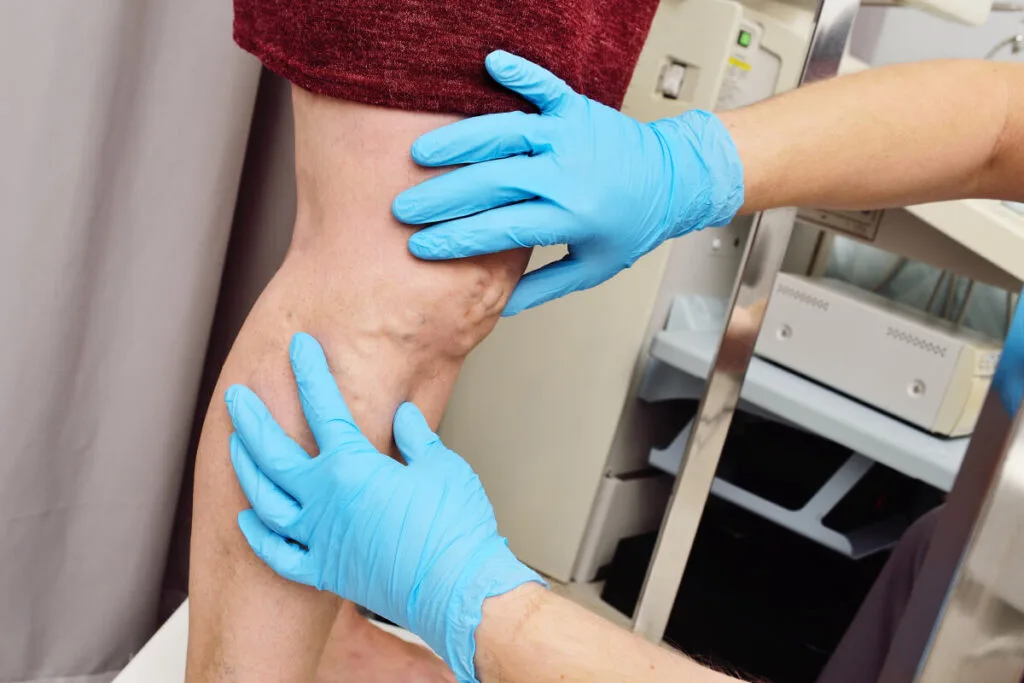
Chronic venous insufficiency may present with dilated leg veins, edema, leg pain, and changes in the skin such as changes in quality and texture.
- Dilated leg veins - Enlargement of the leg veins may be classified based on their sizes such as telangiectasias (also known as spider veins, < 3 mm in size), reticular veins (1 mm to 3 mm), and varicose veins (> 3 mm).
- Edema - Edema is a condition characterized by swelling caused by fluid retention. In the lower extremities, it starts in the perimalleolar region and extends up to the legs.
- Leg discomfort - Leg discomfort is the feeling of pain or heaviness after prolonged standing that may be relieved when the legs are elevated at a level above the heart. This may be due to increased pressure and volume in the venous system.
- Hyperpigmentation - The characteristic darkening of the skin of the legs affected by CVI may be due to the build-up of hemosiderin (iron deposits in the tissues) and pronounced eczematous dermatitis (redness and itchiness of the skin associated with CVI).
- Lipodermatosclerosis - This characteristic condition associated with CVI is caused by inflammatory responses in the dermis and subcutaneous tissues.
Similarly, CVI may also cause signs and symptoms that may be directly or indirectly attributed to its pathophysiology. These include:
- Formation of ulcers - Ulcers are chronic, non-healing wounds caused by skin breakdown due to the poor oxygenation and blood flow of the skin. Types of ulcers include venous ulcer (brought by impediments in the blood flow caused by incompetent venous valves or other causes of venous reflux), arterial ulcer (caused by arterial insufficiency), neuropathic ulcer (caused by conditions such as diabetes), pressure ulcer (caused by unrelieved pressure), among others. The promotion of ulcer healing involves addressing its underlying cause such as CVI.
- Anxiety and Depression - Anxiety and depression are psychological conditions indirectly associated with CVI, in that they may be caused by the aesthetic changes associated with CVI.
How Chronic Venous Insufficiency Is Diagnosed
A critical part of treating a disease is establishing its correct diagnosis. Chronic venous insufficiency is no exception to this rule and various techniques have been developed for its correct diagnosis. Diagnostic procedures involved in establishing that a patient has CVI include medical history and physical examination, non-invasive testing procedures, and invasive testing procedures as well.
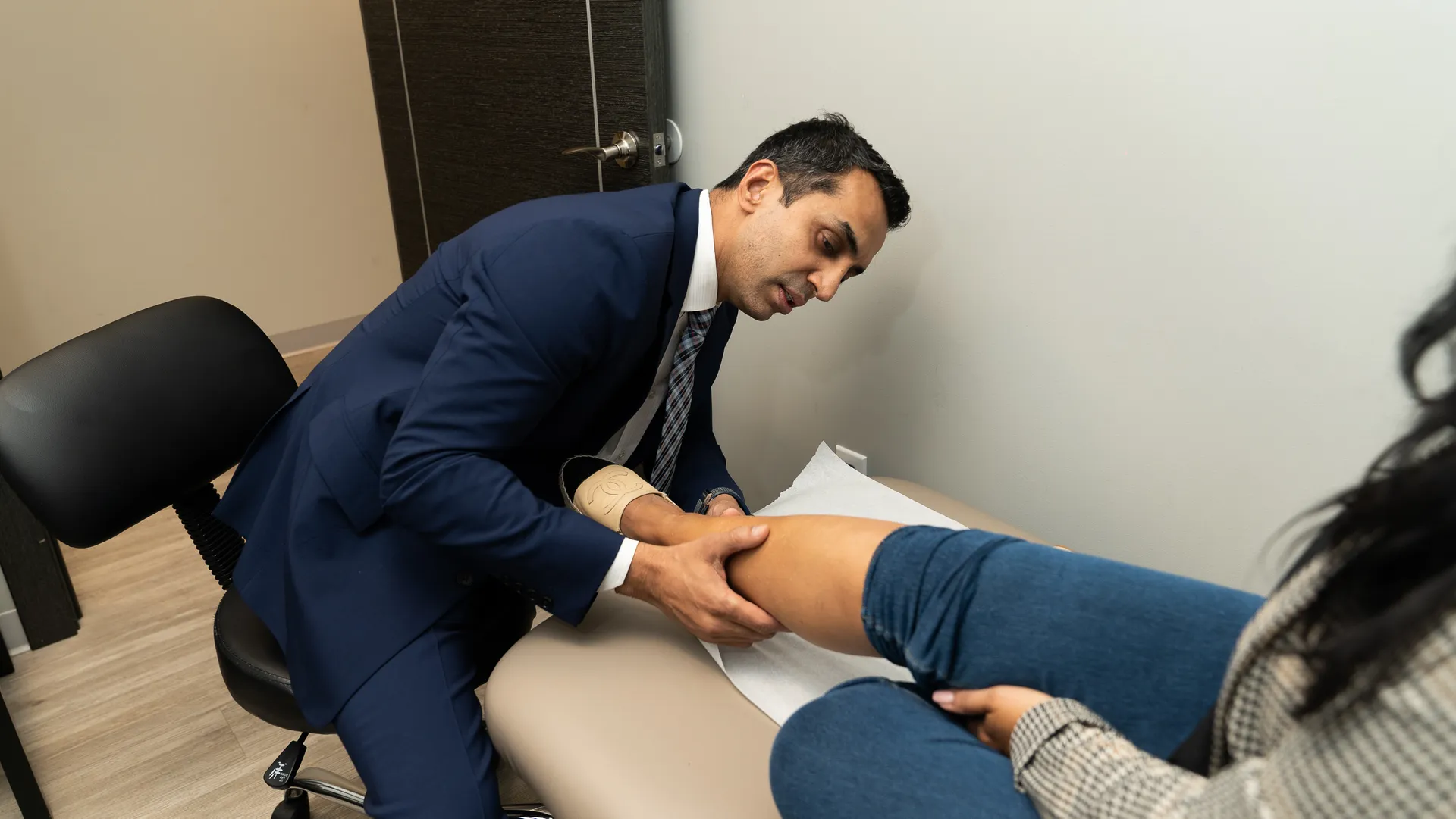
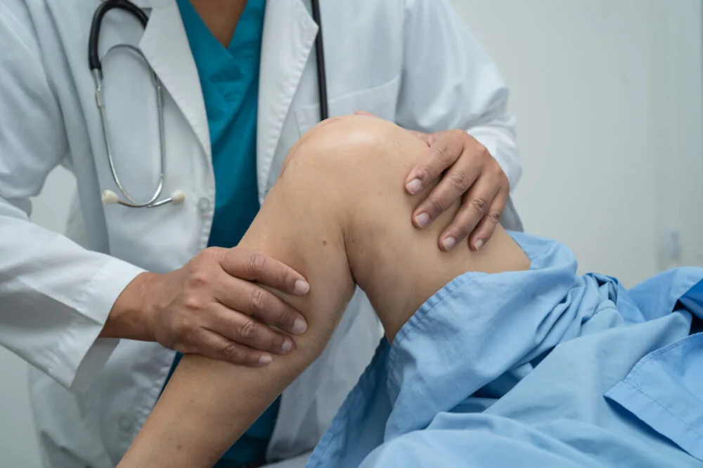
1) Medical History And Physical Examination
History-taking is an important aspect of diagnosing chronic venous insufficiency as it helps determine the risk factors that predispose the patient into developing and exacerbating the disease. On the other hand, physical examination is an important initial assessment that helps look for the signs and symptoms of CVI present in the patient, through examining a patient in an upright position for the maximum distention of veins.
2) Non-Invasive Testing
Non-invasive testing procedures are necessary to determine the involvement and extent of CVI without inserting instruments inside the body. This helps establish the diagnosis of CVI, employing sound waves or air displacement for the visualization of the structures of the body. Examples of diagnostic modalities under non-invasive testing include:
- venous duplex imaging (the most common technique used for the diagnosis of CVI)
- air plethysmography
- computed tomographic
- magnetic resonance venography
- photoplethysmography
- strain gauge plethysmography
- foot volumetry
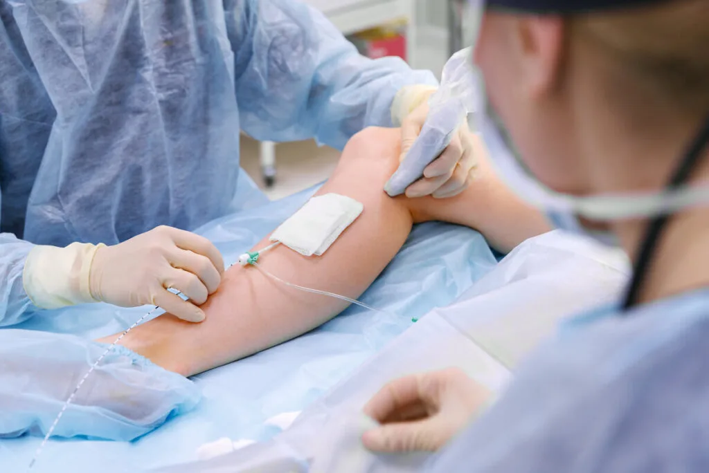
3) Invasive Testing
Invasive procedures involve the injection of contrasts or insertion of instruments such as probes to visualize the vein and help guide interventions. They provide the benefit of improving the detection of the abnormalities of the veins, although they also have risks associated with being invasive such as dissection, perforation, and vasospasm. Examples of diagnostic modalities under invasive testing include:
- contrast venography
- intravascular ultrasound
- ambulatory venous pressure (the gold standard in determining blood mechanisms of CVI)
Vein Center Doctor: Offering Excellent Outpatient Vein Treatments
With our wide range of medical procedures to choose from, we at Vein Center Doctor commit to providing adequate treatment to make our patients healthier. Therapeutic options available in our clinic include radiofrequency ablation, endovenous laser treatment, VenaSeal, and compression therapy.
1) Radiofrequency Ablation
Radiofrequency ablation is an FDA-approved thermal ablation technique which is one of the available minimally-invasive treatments used for treatment of chronic venous insufficiency. It uses a catheter with a heating tip to deliver radiofrequency to the target incompetent vein in order to destroy its inner lining and close it off. This, in turn, leads to the redistribution of blood flow to a healthy vein.
Bruising and tenderness are the possible side effects of this procedure, although this may be avoided by using valvular compression stockings or bandages in the affected leg. It requires minimal downtime and the patients can return to their normal activities shortly.
Caution must also be observed by patients whose superficial veins have a diameter of less than 2 mm and patients who have other medical conditions such as deep vein thrombosis and malignancy as they're advised not to undergo this procedure.
2) Endovenous Laser Treatment
Endovenous laser treatment is another minimally-invasive procedure that uses heat energy to prevent the reflux of the saphenous vein, superficial vein, and perforator vein. It also uses local anesthesia to help the patients not feel pain during the treatment.
Despite the similarity of the principles with radiofrequency ablation, it differs from the said procedure in that this endovenous treatment delivers a laser fiber with wavelengths of 810-nm or 940-nm through its catheter with a heating tip to also close off the blood vessels by destroying their inner lining and formation of scar tissue.
3) Sclerotherapy
Sclerotherapy is a form of treatment that uses sclerosing agents such as 23.4% sodium chloride, sodium tetradecyl sulfate, and sodium iodide to treat many types of venous disease such as varicose veins, bleeding varicosities, and small cavernous hemangiomas.
It has 2 types, liquid sclerotherapy and ultrasound-guided foam sclerotherapy, which, when compared, have the same safety profiles: they have the same side effects namely local inflammation, thrombophlebitis, and skin discoloration, but differ in efficacy in that ultrasound-guided foam sclerotherapy is more effective. This is because it's delivered undiluted to the target veins, unlike liquid sclerotherapy where the sclerosing agent is diluted by blood.
4) VenaSeal
The VenaSeal closure system is a cutting-edge technology that effectively manages vascular diseases without the risk and complications of surgical procedures. This procedure entails the delivery of medical-grade VenaSeal adhesive guided by venous ultrasound. It uses a delivery catheter in a specific delivery sequence where 3 cm of adhesive is delivered every 3 seconds until all vein segments have been provided. The affected area will then be compressed followed by bandaging of the insertion site.
5) Compression Therapy
Compression therapy is a nonsurgical treatment that is the standard of care for chronic venous insufficiency and venous leg ulcer. Adequate compression therapy helps mitigate fluid retention due to venous hypertension, leading to an improved quality of life for compliant patients who can experience improvements in pain, swelling, skin pigmentation, and activity. It's also the standard of care for venous leg ulcers as it's capable of healing and preventing venous ulcers from recurring.
Get Professional Outpatient Vein Treatments At Vein Center Doctor
Venous valvular incompetence is a condition where damage to a venous valve leads to its abnormal functioning. This pathological mechanism, in turn, leads to chronic venous insufficiency and in fact, is a major prognostic indicator as to the progression of the disease state.
At Vein Center Doctor, we're a team of healthcare professionals headed by Dr. Rahul Sood, who are experts in providing venous care with excellent clinical outcomes. We're committed to providing quality patient-centered care through the treatment options that we offer. Start your journey now towards healthier veins by contacting us at 862-227-1143 (NJ) or (862) 500-4747 (NY) and have your free consultation today.