Learning about the possible factors that may predispose a patient to develop a certain disease helps healthcare professionals design measures to employ for prevention of the occurrence of that particular disease. This includes chronic venous insufficiency which is said to be caused by multiple risk factors like diabetes mellitus.
So can diabetes mellitus actually cause chronic venous insufficiency? Yes, because diabetes mellitus can predispose an individual to develop different vascular diseases. This is through different mechanisms like vasoconstriction, inflammation, and thrombosis precipitated by high blood sugar, free fatty acids, and insulin resistance.
The Link Between Diabetes Mellitus and Chronic Venous Insufficiency
Diabetes is considered to be a risk factor for the development of chronic venous insufficiency. In fact, a study has shown that diabetic patients are more likely to develop chronic venous insufficiency, especially when:
- they’re older
- with reduced compliance to treatment
- are also exhibiting peripheral arterial disease
Diabetes also brings in different metabolic abnormalities like hyperglycemia (or the increased levels of blood sugar), increased levels of circulating free fatty acids, and insulin resistance. These abnormalities in turn provoke molecular mechanisms like oxidative stress, protein kinase C activation, and RAGE activation.
These molecular mechanisms promote changes in the function and structure of blood vessels such as the following:
- Vasoconstriction – Vasoconstriction occurs due to the production of mediators that promote this process, such as prostanoids and endothelin. Decreased production of NO, which is a vasodilator, also occurs.
- Inflammation – Increased production of inflammatory mediators such as chemokines, cytokines, and CAMS also occur.
- Thrombosis – Impairments in platelet function in diabetes increase the propensity of the platelets to clump together, favoring the development of venous thrombosis.
Moreover, aside from diabetes, other known risk factors for chronic venous insufficiency include the following:
- Increasing age
- Family history of cardiovascular disease and venous disorders like varicose veins
- High body mass index
- Pregnancy
- Phlebitis
- Previous trauma to the legs
Complications of Chronic Venous Insufficiency
Chronic venous insufficiency, especially when left untreated, may eventually develop into the following complications associated with this disease entity:
- Superficial thrombophlebitis – Superficial thrombophlebitis describes the medical condition in which the veins immediately under the skin (superficial veins) are inflamed and with blood clot formation. This condition is typically not serious, although it may cause severe pain to the patient.
- Venous thromboembolism – Venous thromboembolism is the medical term encompassing deep vein thrombosis and pulmonary embolism. This describes the pathological process in which blood clots are formed in the deep veins, which may be dislodged and reach the lungs.
- Lower extremity ulcers – Lower extremity ulcers are non-healing, chronic wounds that result from impairments in the flow of blood to the skin due to chronic venous insufficiency. This is because less oxygen and nutrients reach the skin.
How Chronic Venous Insufficiency Occurs
The specific etiology of chronic venous insufficiency is yet to be known, but scientists speculate 4 mechanisms in which it occurs, namely hemodynamic and microcirculatory alterations, inflammation, the hypoxic environment, and venous wall remodeling.
1) Hemodynamic and Microcirculatory Alterations
- Valves Competence – Valves are structures in the vein that allows unidirectional blood flow in the vein. This allows blood to go back to the heart despite the low pressure in the veins. The functional status of the valves is important in maintaining this unidirectional blood flow, and impairments in this function result in the backflow of blood and pooling of blood in the veins.
- Calf Muscle Pump Function – Contraction of calf muscles that surround the leg veins such as gastrocnemius and soleus further helps in the return of blood into the heart. The weakening of these muscles results in venous reflux, swelling, chronic venous insufficiency, and, when remaining untreated, in venous leg ulcers.
- Venous Wall Integrity and Elasticity – Veins differ in layer composition from arteries, which makes the veins have less elasticity than arteries but higher extensibility. It’s still uncertain whether this property contributes to chronic venous insufficiency, although there are speculations that veins become stiffer and lose extensibility due to venous thrombosis.
Chronic venous insufficiency also is a result of microcirculatory alterations, which manifest as a decreased number of capillaries, increased permeability of capillaries, and abnormalities in lymphatic microcirculation.
2) Inflammation
Inflammatory cells like neutrophils and T-cells are important mediators in the inflammatory response occurring in the venous wall of patients with chronic venous insufficiency. In fact, these inflammatory cells are found to be in high numbers in these patients compared to normal individuals.
3) The Hypoxic Environment
Hypoxia is the reduced oxygen levels in the tissue. This occurs in the vascular wall of patients with chronic venous insufficiency. Mechanisms of hypoxia in patients with chronic venous insufficiency include the following:
- Endoluminal hypoxia – In endoluminal hypoxia, the endothelium and inner vein wall layers have low oxygen levels due to impediments in blood flow.
- Medial hypoxia – In medial hypoxia, the medial and outer layers of veins have low oxygen levels due to the compression of the vasa vasorum brought by dilation and increased pressure in the veins.
4) Venous Wall Remodeling
Due to shear stress brought by the previously described mechanisms, especially the endothelial changes, a change in the functioning of the body ensues as a response to the alteration in the environment on which the mediators thrive on. These mediators include acidic fibroblast growth factor (aFGF), vascular endothelial growth factor (VEGF), and insulin-like growth factor-1 (IGF-1).
Signs and Symptoms of Chronic Venous Insufficiency
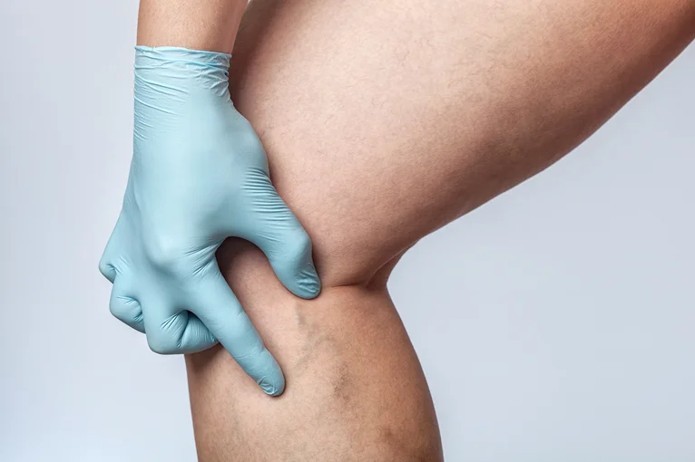
Common clinical manifestations of chronic venous insufficiency extend not just to the veins and muscles but to the skin as well. These signs and symptoms include the following:
- Dilated veins – Dilated veins may be classified into 3, which may be differentiated based on their diameter size. Telangiectasias or spider veins occur in veins less than 1 mm, reticular veins are 1 to 3 mm in diameter, and varicose veins are more than 3 mm in diameter.
- Venous Edema – Venous edema defines the condition in which increased volume of fluid in the skin and subcutaneous tissue results in a characteristic swelling observable in the legs. It usually occurs in the ankle but may also be seen extending up to the leg and foot.
- Subjective symptoms – Subjective symptoms associated with chronic venous insufficiency include leg pain, heaviness, and fatigue. Sensations of swelling, cramps, itching, and tingling may also be felt. The restless leg syndrome may also be felt.
- Skin changes – Typical skin changes associated with chronic venous insufficiency include hyperpigmentation of the affected skin, erythematous dermatitis, lipodermatosclerosis or the localized chronic inflammation of the skin and subcutaneous tissue, and atrophie blanche or white atrophy.
How Chronic Venous Insufficiency is Diagnosed
Chronic venous insufficiency may be diagnosed first by careful history taking where risk factors for this vascular disease are ascertained. This is followed by a physical examination where an inspection of the signs and symptoms of chronic venous insufficiency is done.
Complementary to history taking and physical examination for the diagnosis of chronic venous insufficiency are imaging methods, which may be non-invasive or invasive:
| Non-Invasive Methods | Invasive Methods |
| Venous Duplex Imaging Air Plethysmography Computed Tomography or Magnetic Resonance Venography Photoplethysmography Strain Gauge Plethysmography Foot Volumetry | Contrast Venography Intravascular Ultrasound Ambulatory Venous Pressure |
How Chronic Venous Insufficiency is Treated
Signs and symptoms associated with chronic venous insufficiency may be treated conservatively or with the use of interventional methods.
1) Conservative Methods
Conservative methods of treating chronic venous insufficiency clinical manifestations involve lifestyle modification like regular exercise to maintain a healthy weight and strengthen calf muscle pump function. It also involves the use of medications for symptomatic relief, such as the following:
- Low-dose diuretics for edema
- Systemic antibiotics (with the prescription of a healthcare professional) for infections like cellulitis and infected stasis ulcers
- Skincare and wound care of patients with impairments in skin integrity like stasis dermatitis and chronic leg ulcer
2) Interventional Methods
Similarly, interventional methods involve outpatient vein treatments and surgical interventions. For vascular surgery, typical methods include the following:
- Venovenous bypass (Palma procedure), endovenous angioplasty, and iliac vein stenting for the treatment of venous obstruction;
- Phlebectomy or vein stripping;
- Subfascial endoscopic perforator surgery;
- Valvuloplasty or the restoration of valve function.
Vein Center Doctor: Your Partner in Managing Chronic Venous Insufficiency
At Vein Center Doctor, we can help you manage your signs and symptoms of chronic venous insufficiency through our expertise in the different outpatient procedures available. We offer radiofrequency ablation, endovenous laser treatment, sclerotherapy, VenaSeal closure system, and compression therapy that are all proven to have good efficacy and safety profiles.
1) Radiofrequency Ablation
Radiofrequency ablation is a minimally-invasive treatment that closes off the target vein using radiofrequency energy. This procedure involves the use of local anesthesia to numb the area to be inserted with a catheter, and ultrasound to visualize its insertion.
The heat-tipped catheter is inserted up to the saphenofemoral junction, delivering radiofrequency energy through an endovenous electrode. This then inflicts injury to the vein wall of the target vein, leading to its closure and the consequent redistribution of blood flow to healthy veins.
This is perfect for varicose veins that have large diameters but can’t be done on small leg veins with a venous disease like spider veins and reticular veins.
2) Endovenous Laser Treatment
Endovenous laser treatment is an interventional procedure that employs the same principle as radiofrequency ablation.
It entails the delivery of laser energy using a heat-tipped catheter to close off the target veins but at a slower rate than radiofrequency ablation. It also uses local anesthesia and ultrasonography.
This is also good for varicose veins that can be catheterized but can’t be done on spider veins and reticular veins. Side effects of this procedure include bruising and hyperpigmentation.
3) Sclerotherapy
Sclerotherapy involves the delivery of chemicals called sclerosing agents to the target vein to irritate its vein wall, effectively resulting in its closure. Typical sclerosing agents used include the hypertonic saline solution and sodium tetradecyl sulfate.
It exists in liquid or foam form, with the foam injections being the more effective but comes with more side effects.
It’s an excellent procedure to be used for spider veins and reticular veins, although it can’t be done for those who are pregnant, breastfeeding, and have known allergies to the sclerosing agents used. Typical complications include hyperpigmentation and deep vein thrombosis.
4) VenaSeal Closure System
The VenaSeal closure system is a minimally-invasive procedure that doesn’t require anesthesia before the procedure and the use of compression garments as adjunctive therapy after the procedure.
This procedure entails the delivery of the VenaSeal adhesive, a special formulation of n-butyl-2-cyanoacrylate, to the target vein using a special catheter. The procedure is done with the aid of ultrasound and guidewire, both of which help in the insertion of the catheter.
The procedure is done first by flushing with saline solution. The VenaSeal adhesive is then delivered in a pattern where 0.1 cc is delivered for 3 seconds, then the area is compressed for 3 minutes. This is repeated until all the target vein is delivered with the VenaSeal adhesive.
5) Compression Therapy
Compression therapy is considered to be the standard-of-care treatment for varicose veins and venous ulcers. It works by opposing the hydrostatic pressure that leads to other complications of chronic venous insufficiency like edema and venous ulcers.
With the use of compression garments or other medical devices like intermitted pneumatic compression pumps, graded pressure is delivered to the desired area. The table below summarizes the level of compression required per complications of chronic venous insufficiency:
| Clinical Manifestation | Tension (mmHg) |
| Varicose veins with or without edema | 20-30 |
| Advanced venous skin changes or ulcers | 30-40 |
| Recurrent ulcers | 40-50 |
Get High-Quality Outpatient Vein Treatments at Vein Center Doctor
There’s an increased risk of developing chronic venous insufficiency if a patient has diabetes mellitus. This is because of the different hemodynamic mechanisms that occur in patients with diabetes mellitus that bring impairments in the functions of blood vessels.
At Vein Center Doctor, we’re committed to bringing improvements in the quality of life of our patients through our high-quality outpatient vein treatments. We complement the newest innovations with the skills of our team headed by Dr. Rahul Sood. Contact us today at 1-862-500-4747 for your free consultation on vein care.


